

Preimplantation genetic testing of embryos (PGT)

Preimplantation genetic testing of embryos (PGT) is an advanced method linked to assisted reproduction techniques that allows embryos to be genetically tested before they are transferred into the uterus, known as embryo transfer. The aim is to select the embryo with the highest chance of healthy development and to reduce the risk of miscarriage or transmission of an inherited disease to future generations.

Preimplantation genetic testing (PGT)

Preimplantation genetic testing of embryos is a modern method combining assisted reproduction with clinical genetics, which makes it possible to detect genetic abnormalities in embryos at an early stage of development and helps reduce the risk of unsuccessful embryo transfer, miscarriage or the birth of a child with a serious genetic disease.

Scroll down
Preimplantation genetic testing of embryos

Principle of IVF treatment with preimplantation genetic testing of embryos

Preimplantation genetic testing of embryos

Cultivation and preparation of the embryo for biopsy

After the eggs are fertilised, the resulting embryos are cultured under strictly controlled laboratory conditions until day 5–6, when, in the optimal case, they develop into the stage of a so-called hatching blastocyst. This is the ideal phase for the safe collection of cells, known as biopsy, intended for preimplantation genetic testing.

Scroll down

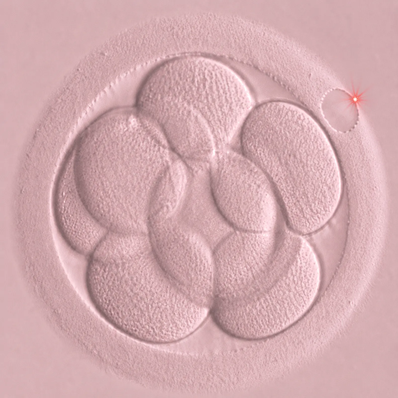
Assisted hatching as part of cultivation

In order to obtain the highest possible number of embryos at this stage, we perform assisted hatching on all embryos on day 3 of culture. This targeted laser procedure supports the natural release of the embryo from its protective shell (zona pellucida) and creates suitable conditions for a gentle and safe biopsy.

Scroll down
Preimplantation genetic testing of embryos

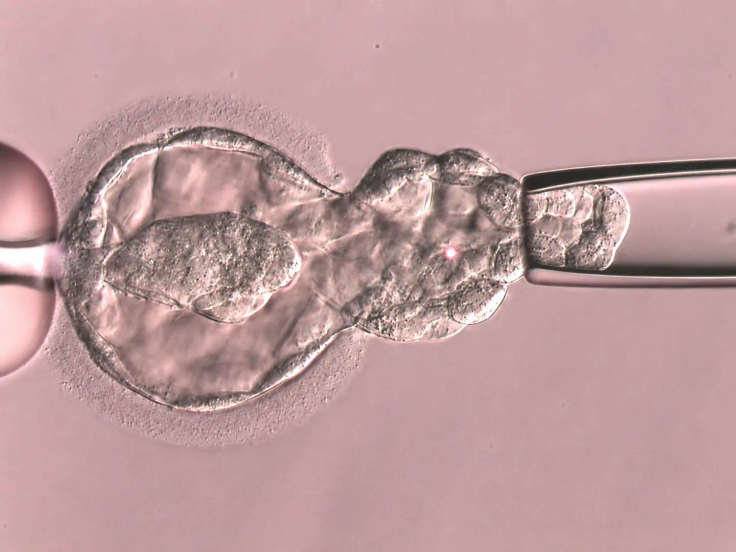
Gentle cell collection for genetic analysis

Biopsy is a laboratory procedure in which 5 to 10 trophectoderm cells are gently collected from the blastocyst using a micropipette and laser. These cells form the basis of the placenta and embryonic membranes, not the embryo itself, known as the embryoblast. The procedure therefore does not negatively affect the further development of the embryo. The collected material is transferred into a microtube and subjected to detailed genetic analysis in a genetic laboratory according to the selected type of genetic test:

Scroll down

After biopsy, the embryos are frozen using the very gentle method of vitrification

Genetic testing of embryos takes 3 to 5 weeks, depending on the type of genetic test. After completion, an embryo in which no genetic abnormalities monitored by the selected method were detected is chosen for frozen embryo transfer (FET/KET).

Steps of genetic analysis

1. DNA preparation from collected cells

From a small number of cells, approximately 5–10, enough DNA is prepared in the laboratory for analysis of the entire embryo genome, meaning all of its genetic information. This step is called whole-genome amplification and makes it possible to perform detailed testing even from a very small embryo sample.

2. Comprehensive and targeted genetic analysis of embryos

Different molecular genetic methods are used for embryo genetic testing depending on the type and purpose of the test. To set them up correctly, it is necessary to have the parents’ karyotypes available, meaning testing of the number and structure of chromosomes, or a genetic report describing the monitored monogenic mutation, meaning a genetic abnormality in one specific gene.

NGS (next-generation sequencing)

NGS (next-generation sequencing) is used for an overall assessment of the embryo's genetic status. It allows the number of chromosomes to be tested and can also detect structural chromosome abnormalities that may affect further embryo development.

Karyomapping

Karyomapping is an indirect method used mainly for monogenic diseases within PGT-M. By comparing DNA reference samples from the parents and other family members, it makes it possible to determine whether the embryo has inherited the monitored genetic disease and also includes chromosome number testing, as in PGT-A.

Direct mutation detection

Direct mutation detection is targeted testing in which a specific mutation variant in the monitored gene is analysed. The most commonly used methods are PCR and sequencing, such as Sanger sequencing, which precisely determine the presence or absence of the given mutation in the embryo. Direct mutation detection is mainly used when it is not possible to obtain DNA reference samples from relatives needed for indirect methods, which are more reliable from a diagnostic perspective.

3. Selection of a genetically healthy embryo

Based on the results, the laboratory recommends which embryo has the highest chance of successful pregnancy and healthy development. In the meantime, the embryos are safely frozen and their transfer takes place only after the results have been evaluated.


Why PGT is useful

It increases the chance of successful transfer and pregnancy.

It reduces the risk of miscarriages caused by genetic errors.

It helps prevent the transmission of inherited diseases.

Types of PGT according to the genetic abnormality

PGT-A

Preimplantation genetic testing for aneuploidy

PGT-A is a genetic test used to assess the correct number of chromosomes in embryo cells before the embryo is transferred into the uterus. The tested karyotypes of the parents are sufficient for performing this test.

Every cell in the human body, except for eggs and sperm, contains 46 chromosomes. If an embryo has an extra chromosome or, conversely, is missing one, known as aneuploidy, this may lead to unsuccessful embryo implantation, recurrent miscarriages or the development of genetically determined conditions, such as Down, Edwards or Patau syndrome.

PGT-A is intended especially for:

  • women of advanced reproductive age, over 35 years,
  • couples with repeated unsuccessful embryo transfers,
  • couples with recurrent miscarriages or the birth of a child with a developmental defect,
  • men with a severe semen analysis disorder, teratozoospermia, after chemotherapy or after surgical sperm retrieval (TESE).

Main benefits of PGT-A

PGT-A makes it possible to detect numerical chromosome abnormalities before embryo transfer and select the embryo with the highest potential for implantation and healthy development, thereby significantly reducing the risk of miscarriage and supporting more effective and targeted infertility treatment.

PGT-A PLUS

Extended preimplantation genetic testing for aneuploidy

PGT-A PLUS is an extended form of preimplantation genetic testing for aneuploidy which, in addition to determining the correct number of chromosomes in the embryo, also provides information about the origin of detected abnormalities.

What PGT-A PLUS offers in addition to standard PGT-A:

  • it determines the parental origin of chromosome abnormalities, meaning whether the genetic abnormality comes from the egg or from the sperm,
  • it helps distinguish whether the genetic error was already present in the reproductive cells, meaning the egg or sperm, or whether it occurred only during early embryo development.

Genetic analysis using a reference DNA sample

Unlike standard PGT-A, which is performed using NGS, meaning next-generation sequencing, PGT-A PLUS is analysed using SNP-array technology, meaning analysis of single nucleotide polymorphisms. This method makes it possible to monitor inherited genetic markers and determine whether the detected chromosomal abnormalities originate from the egg or the sperm. Therefore, in addition to karyotypes, a reference DNA sample from one of the parents is required for the test, as it serves to precisely clarify the origin of the genetic change.

Main benefit of PGT-A PLUS

The PGT-A PLUS method provides extended genetic information, especially for couples with repeated treatment failures, and supports an individualised approach to treatment. It helps better understand the origin of detected genetic abnormalities and, based on this information, targetedly adjust the next treatment steps, for example by considering the use of donated eggs or sperm in a subsequent cycle.

PGT-SR

Preimplantation genetic testing for structural rearrangements

PGT-SR is genetic testing of embryos focused on detecting structural chromosome abnormalities in which parts of chromosomes are exchanged, moved or lost. The aim is to select an embryo with a complete and correctly arranged karyotype that has the highest chance of successful implantation and healthy development.

PGT-SR is intended for couples in whom a balanced structural chromosomal abnormality has been detected in one partner, such as a translocation or inversion, which does not affect their health, but does affect their reproduction. These abnormalities lead to a higher formation of reproductive cells (eggs/sperm) with an unbalanced rearrangement, which causes embryos to develop with unbalanced genetic information, where part of the genetic information is missing or present in excess. Such embryos have a higher risk of spontaneous miscarriage or congenital developmental defects.


Genetic analysis based on karyotype

PGT-SR uses the NGS method, meaning next-generation sequencing, to detect structural chromosome rearrangements in embryos. For genetic analysis, the karyotype of the parent with the precisely identified abnormality is essential, as embryo testing is then targeted to this abnormality.

Main benefits of PGT-SR

PGT-SR makes it possible to select an embryo without structural chromosomal abnormalities, reduces the risk of unsuccessful pregnancy and miscarriage, and supports a targeted and individualised approach to infertility treatment.

PGT-M

Preimplantation genetic testing for monogenic diseases

PGT-M is the most detailed genetic test of embryos and is used to detect specific inherited diseases caused by a change, meaning a mutation, in a single gene. This method makes it possible to select embryos that do not carry the monitored genetic disease before they are transferred into the uterus.

PGT-M is intended for couples:

  • in whom a genetic disease is known in the family, for example mutations associated with a high risk of cancer development – BRCA1, BRCA2, Huntington's disease or myotonic dystrophy,
  • where the partners are carriers of an inherited disease, such as cystic fibrosis, spinal muscular atrophy or phenylketonuria,
  • who have an increased risk of passing a specific genetic mutation on to their child, for example the risk of recurrence of a randomly occurring mutation in another child, such as neurofibromatosis.

Genetic analysis using karyomapping with reference DNA samples

The collected embryonic cells are genetically tested using the karyomapping method, which makes it possible to indirectly and very precisely determine whether the embryo carries a specific inherited mutation. The test is based on comparison of so-called genetic markers (SNPs – single nucleotide polymorphisms), meaning markers located around the monitored gene, with reference DNA samples from the parents and another affected/healthy family member.

These reference samples serve to create an individual genetic "map" of the family. Thanks to this, it is possible to reliably determine whether the embryo has inherited the healthy or mutated variant of the gene without having to analyse the gene itself using so-called direct detection. This indirect approach also significantly reduces the risk of so-called allelic dropout (ADO), meaning a situation in which one of the parental genetic variants is not detected during genetic analysis, which could lead to a false-negative result.

Allelic drop-out (ADO) occurs due to the analysis of a very small amount of DNA obtained from a limited number of embryo cells. During laboratory amplification, meaning DNA multiplication, preferential amplification of only one genetic variant may occur, meaning genetic information from only one parent, while the genetic information from the other parent remains undetected.

The karyomapping method therefore provides high accuracy and reliability of genetic testing. Another advantage is that it simultaneously assesses the number of chromosomes in the embryo, as in PGT-A, which makes it possible to select embryos with the highest potential for healthy development and successful pregnancy.

Main benefits of PGT-M

PGT-M provides an individually targeted approach to infertility treatment that makes it possible to select and transfer embryos without the monitored genetic mutation and helps prevent the transmission of a serious inherited disease to future generations.

FAQ

Does cell collection affect embryo development?

No. Only a few cells are collected from the embryo's trophectoderm, the part of the embryo from which the placenta later develops, at the stage of a so-called hatching blastocyst.

How long does genetic analysis take?

Processing and testing the embryonic DNA sample in the genetic laboratory takes approximately 3 to 5 weeks, depending on the selected type of genetic test.

Does PGT-A increase the chance of pregnancy?

Yes. Selecting an embryo with the correct number of chromosomes increases the chance of successful implantation in the uterus and reduces the risk of miscarriage.

Decorative graphic
Donation coordinator

Mgr. Hana Ströer

IVF Coordinator

Hlinky 144, 603 00 Brno, CZ

Do you need more information?
Contact us!

Potřebujete více informací?
Ozvěte se nám!